New Australian research has outlined the first radiation reference levels for digital breast tomosynthesis mammography.
Compressed breast thickness – not breast density – is more closely associated with radiation dose in breast imaging, new research suggests.
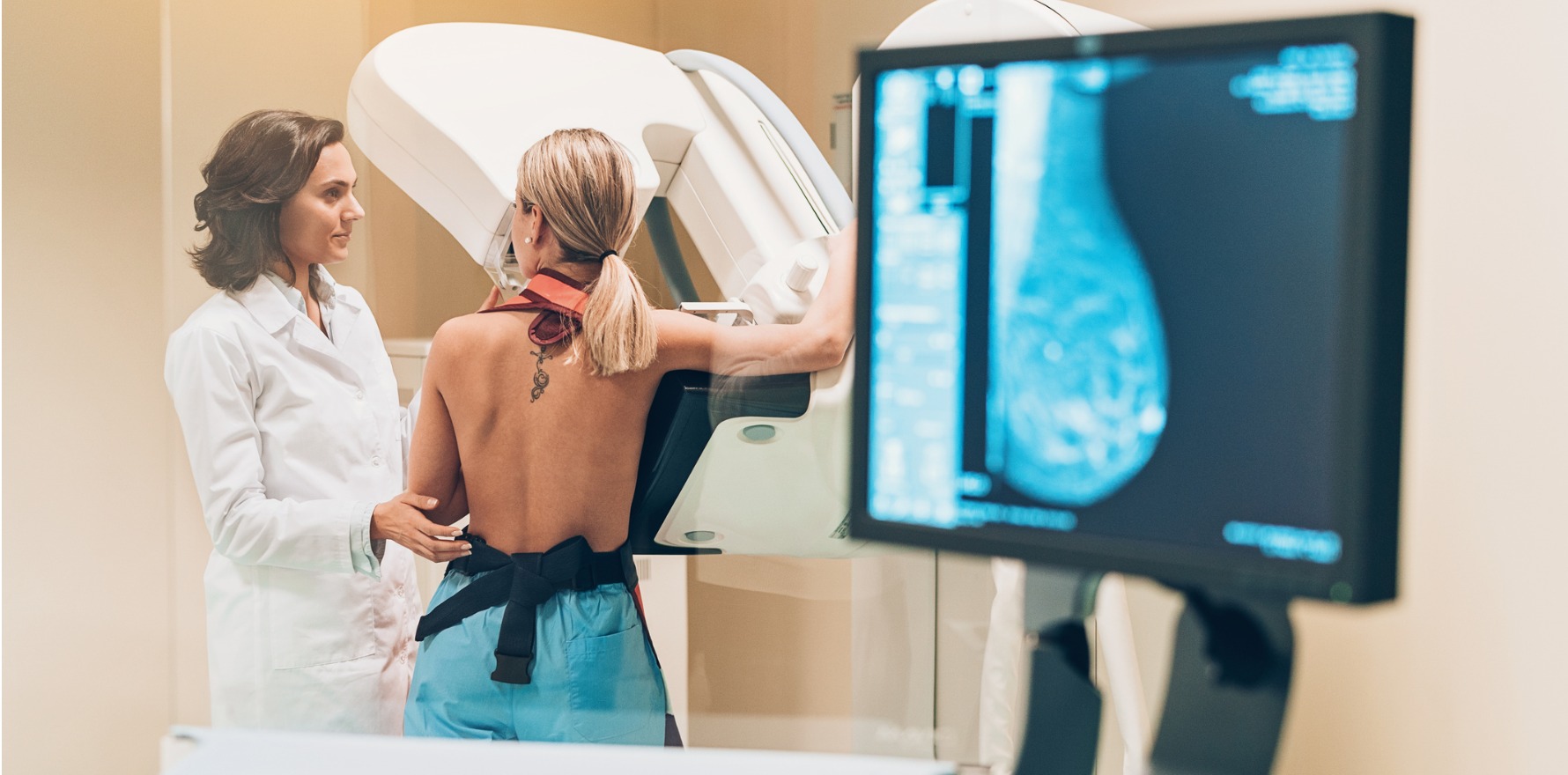
Digital breast tomosynthesis mammography is the recommended diagnostic imaging approach for people with a higher risk of developing breast cancer or who are already showing symptoms of breast cancer.
However, there are no national guidelines that set out the amount of radiation that should be used in this imaging technique.
As a first step to establishing diagnostic reference levels for DBT, a team of Victorian radiographers identified that the radiation dose used in DBT was closely related to compressed breast thickness – with thicker breasts receiving higher radiation doses. Their findings were published in the Journal of Medical Radiation Sciences.
“We know breast tissue is particularly radiosensitive. Tracking the radiation dose patients receive during DBT scans is really important for quality assurance and patient safety,” mammographer and board director of the Australian Society for Medical Imaging and Radiation Therapy Bianca Magill told media.
“This research represents the first step towards diagnostic reference levels. More studies contributing further data and analysis will lead to important standards in Australian mammography.”
The researchers analysed data from over 9000 mammographic images collected between November 2019 and December 2022. The cohort of included patients undergoing DBT during this timeframe ranged from 29 to 90 years, with a median age of 59 years.
The majority of patients were classified as having scatted fibroglandular tissue or heterogeneously dense breasts (categories B and C on the Breast Image-Reporting and Data System density classification; approximately one third of the entire cohort each). The patients were split between density A (almost entirely fatty) and density D (extremely dense).
The level of radiation used for each image varied between 0.72 and 5.94mGy (median average glandular dose 2.23mGy). Regardless of how dense the patients’ breast was, the average glandular radiation dose displayed a strong positive correlation with compressed breast thickness, with higher radiation doses associated with thicker breasts.
Patients were divided into one of four groups based on their compressed breast thickness, and local diagnostic reference levels were established from the median and 75th percentile of the radiation doses delivered during DBT for each patient.
The radiation dose benchmarks for each compressed breast thickness group were: 1.5mGy for 13-49mm, 2.70mGy for 50-74mm, 3.90mGy for 75-99mm, and 4.70mGy for 100-118mm.
“The correlation between average glandular dose and compressed breast thickness implies that the thickness to which the mammographer compresses the breast tissue is an important factor to determine average glandular dose,” the researchers wrote.
“For any given thickness, optimal compression will result in a shorter exposure time and equalisation of breast thickness which may reduce breast dose. Compression force applied during a DBT examination is operator and patient dependent, which impacts CBT and ultimately dose.
“This patient-based study will serve to assist in creating simple local diagnostic reference level guidelines and values for a range of compressed breast thicknesses for Australians to optimise DBT procedures.”
Related


DBT is a different type of mammography that uses 3D imaging, unlike breast cancer screening which typically uses 2D imaging. An x-ray tube moves in an arc around the breast and uses a small dose of ionising radiation to image the breast tissue.
Diagnostic reference levels – which provide an idea of whether the amount of radiation administered during a routine medical procedure is abnormally high or low – are published by the Australian Radiation Protection and Nuclear Safety Agency in Australia.
Reference levels exist for other diagnostic imaging approaches, such as CT/PET scans, nuclear imaging and coronary angiography.
